Cells are the structural and functional units that cover the entire human body. Instead of the cells having an individual structure, they are grouped into clusters. We call these clusters ‘tissues’. Biologically, tissues are groups of cells that have a collective function. The cells might be structurally different and have separate functions. Nevertheless, as a whole, the cells make the work of the entire tissue as a single function. Their functions collectively are responsible for the same.
The origin of the word ’tissue’ is from the French word ’tissue.’ ‘Tissue’ means ‘woven.’ Therefore, a tissue is a cluster of cells ‘woven’ together to function as a unit.
Histology is the study of human or animal tissues. While histopathology refers to the study of tissues in case of diseases or mutations. A tissue is a level of organization between the individual cells and the completely developed organs. A tissue is a combination of similar cells and their extracellular matrix from the same origin. They together carry out a specific function. Multiple tissues functionally combine to create an organ.
The diagram below shows the different tissues located within the body.

Tissue Types
The human body has mainly four types of tissues distributed within the body.
The types are as follows;
- EPITHELIAL TISSUE
- Epithelial tissue is the group of cells that cover the outer surfaces of the body. Most of the epithelial tissue is located towards the outer surface of the body. Their origin is ectodermal, and they usually protect the body from external agents or irritation. They also help us sense various things through our surroundings.
- Examples include skin epidermis etc.
- CONNECTIVE TISSUE
- As the name implies, connective tissue connects various segments of the body. They bind the organs of the body together. They usually have multiple functions of variety that conduct internal processes in the body.
- Examples include blood, bones, areolar tissue, adipose tissue, etc.
- MUSCULAR TISSUE
- Muscular tissue constitutes the muscles in the body. Within the body, muscular tissues have multiple functions. The functions can include movement, peristalsis, and even heart pumping. Muscular tissues have a complex structure and are of multiple types.
- NERVOUS TISSUE
- Nervous tissues constitute all the tissues in the body that create our nervous system. Their main task is sensation and transmission of responses throughout the body. Nervous tissues mostly do not multiply after birth.
- Examples include nerves, microglia, etc.

Connective Tissue – Types And Functions
Connective tissue is the group of tissues in the body that maintain the structure of the body and its organs. They also provide cohesion and internal support. The connective tissues include different types of fibrous tissue that vary only in their density and the number of cells present within them, as well as the more specialized and identifiable types.
In the cavity of the abdomen, most organs are suspended from the abdominal wall by a special membranous band called the mesentery. In such tissues, the cells specialize in the production and preservation of fat or lipids.
The primary supportive structure of the entire body from within is formed by a skeleton composed of bone, a type of connective tissue that has great resistance to stress owing to its highly organized laminated structure and to its hardness, which results from the deposition of mineral salts such as Calcium in its fibers and amorphous ground matrix.
Read Also: Deletion Mutation: Definition, Examples & Diseases
Blood is also a specialized connective tissue. It is among the very few liquid tissues. Blood is composed of specialized cells of different kinds. These cells form the cellular component of blood. While the liquid matrix of blood is referred to as plasma. Plasma acts as a reaction medium for the corpuscles. Human blood is mildly alkaline.
Areolar tissue is a kind of packing tissue that is also a type of connective tissue. The packing of such tissue is loose. The matrix is not hard either. Henceforth, areolar tissue is classified under loose connective tissues.

Loose Connective Tissue
Biologically, the loose connective tissue is a type of connective tissue which consists of areolar tissue, reticular tissue, and adipose tissue. Loose connective tissue is the most widespread type of connective tissue that occurs in vertebrates. It holds the body organs in position and attaches the covering epithelia to other surrounding tissues.
For example, it surrounds the nerves and blood vessels. Fibroblasts are specific cells widely spread out in this tissue. Fibroblasts are irregular branching cells that secrete strong fibrous proteins and proteoglycans as an extracellular matrix surrounding the cells. The cells of areolar connective tissue are generally separated by a considerable distance by a pliable gelatinous substance primarily made up of collagen and elastin.
The nomenclature of loose connective tissue is based on the “weave” and the type of fibers within it. There are three main types;
- Collagenous fibers –
- Collagenous fibers consist of collagen. They consist of bundles of fibrils that are coils of collagen molecules.
- Elastic fibers –
- Elastic fibers consist of elastin protein and are “stretchable.”
- Reticular fibers –
- Reticular fibers consist of one or more types of very thin collagen fibrils. They have the function of joining connective tissues to other tissues.

Areolar Tissue
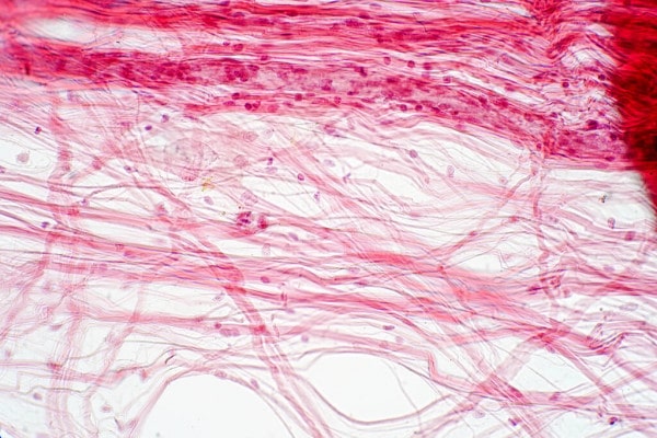
Areolar tissue is the most widely distributed solid connective tissue in the body. Which makes sense because areolar tissue is present under the entire skin.
And areolar tissue also supports the epithelium. Areolar tissue contains randomly spread out fibers, fibroblasts, mast cells, and macrophages. The areolar tissue supports the various organs located in the gut cavity. Areolar tissue also fills the space between muscle fibers and wraps around blood and lymph vessels.
Areolar Tissue Functions
Its functions of areolar tissue are as follows:
- Areolar tissue supports the internal organs
- Such tissue assists in the repair of tissues of muscles and skin also
- Areolar tissue helps in packaging between the organs by filling the space inside the organs
Therefore, now we can delve into the structure of the tissue.
Areolar Tissue Structure
Structurally, areolar tissue has a viscous, gelatinous consistency, which may fluctuate in different parts of the body due to changes in temperature or pH. Alongside this, areolar tissue allows gliding between various muscles and organs. And areolar tissue also permits the diffusion of substances like Oxygen or nutrients from small vessels to the cells. Henceforth, also aiding the diffusion of metabolites back to the vessels.
Areolar Tissue Location
The areolar tissue is located beneath the layer of the dermis. Areolar tissue is also present underneath the epithelial tissue of all the body systems that have external openings. This tissue is widely located within the body and helps in protection and cushioning primarily.
Areolar tissue is the initial site where antigens and other bacteria or pathogens, and other agents that have breached an epithelial surface can be killed. It also forms a mesh-like tissue with a fluid matrix that supports the epithelial tissues, such as the skin and other covering membranes.
Areolar tissue fills the spaces between various organs. Thereby holding them in place while providing them with protection and cushioning.